返回

使用波形识别不同步 - 步骤 2

文章

作者: Caroline Brown, Branka Cupic

日期: 30.09.2022

在上一期中,我们的床旁建议介绍了使用呼吸机波形识别不同步的起点。

使用波形识别不同步 - 步骤 2

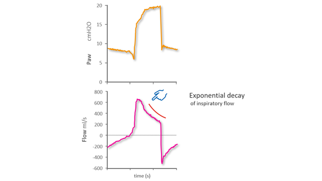
第一步是了解正常的呼吸是什么样的,以及如何识别病人和呼吸机之间的良好同步性。我们介绍了指数递减的概念,以及如何识别吸气努力的开始和良好同步的吸气结束。

下一步是如何识别最常见的较小和较大不同步。

在更仔细地介绍这些之前,我们将回顾构成 Mojoli et al 应用的波形分析系统方法基础的原理。 (Mojoli F, Pozzi M, Orlando A, et al. Timing of inspiratory muscle activity detected from airway pressure and flow during pressure support ventilation: the waveform method.Crit Care.2022;26(1):32.Published 2022 Jan 30. doi:10.1186/s13054-022-03895-41​)。

  • 正常呼吸形式的病人吸气是主动的,呼气是被动的
  • 流量指数递减表示被动状况(适用于吸气和呼气流量)
  • 在同步压力支持通气情况下,在呼吸机呼气阶段应只观察到被动状况
  • 呼吸机吸气阶段的被动状况表示自动触发或延迟切换
  • 呼吸机呼气阶段的被动状况偏差表示提早切换、无效吸气努力或呼吸肌激活

较小的不同步 - 提早切换

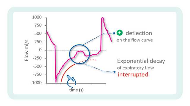
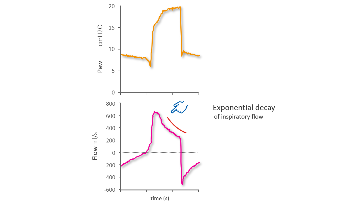
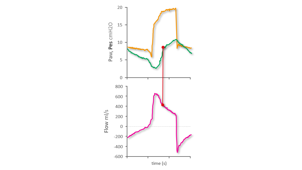
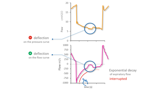
如果病人和呼吸机是同步的,并且切换是最佳的,流量波形将显示呼气开始时的峰值呼气流量,然后是指数递减。提早切换(即呼气在吸气努力结束之前开始),用初始呼气流量的变形来表示:正常的峰值流量被向上偏移所取代,而正常的指数递减开始较晚(见下图 1)。

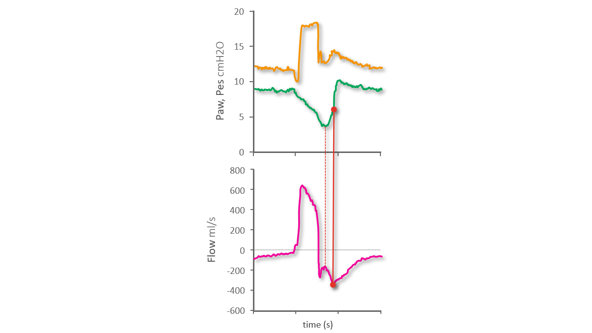
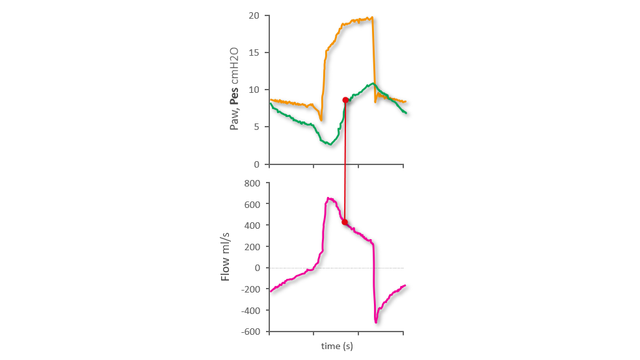
使用食道压 (Pes) 波形作为参考,我们可以看到食道压的最低点(即最大吸气力的点)发生在呼气期间,并与呼气流量的向上偏移相对应。指数流量递减开始较晚,即在吸气努力结束时,如食道压波形上所示(见下图 2)从我们上一个床旁建议中,我们知道食道压在最低点后的快速增加表示吸气肌肉放松,而这个快速增加的中点可以作为吸气结束的参考。

显示向上偏移的波形
图 1(图像来自 Mojoli et al.Critical Care (2022) 26:32)
显示向上偏移的波形
图 1(图像来自 Mojoli et al.Critical Care (2022) 26:32)
显示指数递减开始较晚的波形
图 2(图像来自 Mojoli et al.Critical Care (2022) 26:32)
显示指数递减开始较晚的波形
图 2(图像来自 Mojoli et al.Critical Care (2022) 26:32)

较小的不同步 - 延迟切换

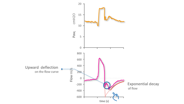
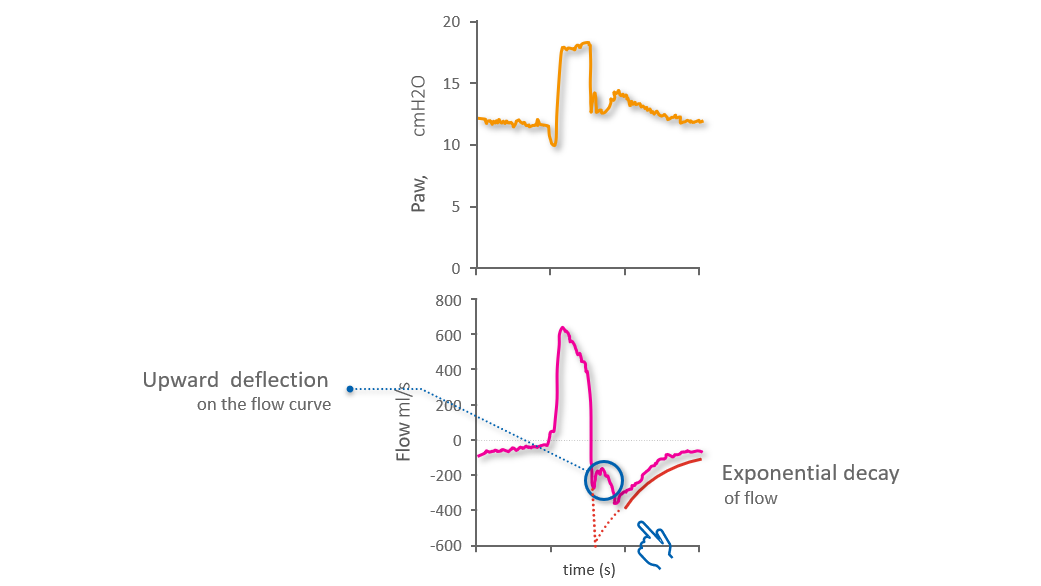
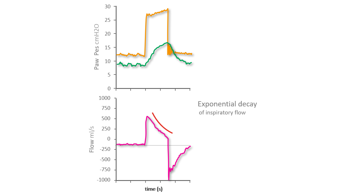
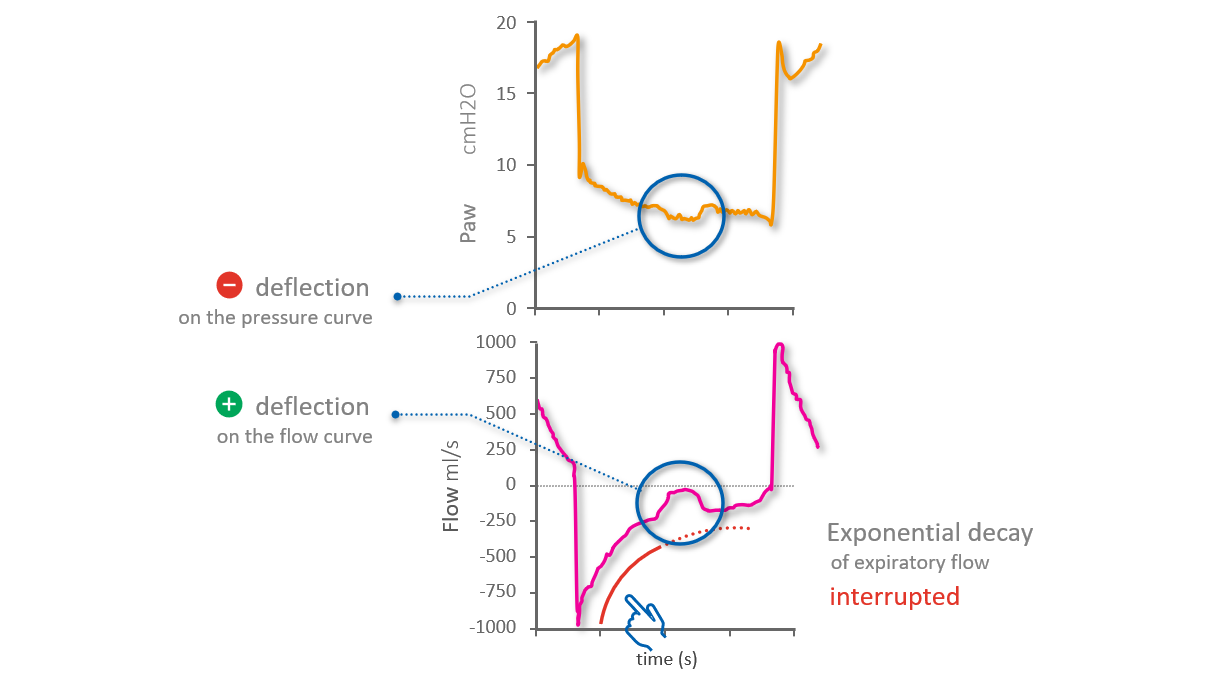
在延迟切换到呼气的情况下,有两个吸气阶段。第一阶段是由吸气肌肉的正常同步拉动和呼吸机的推动引起的,但吸气随后仅由于呼吸机的推动而继续。从而导致被动吸气。您可以从吸气流量在第一阶段后出现的向上凸起的长时间指数递减中在流量波形上识别出这一点(见下图 3)。
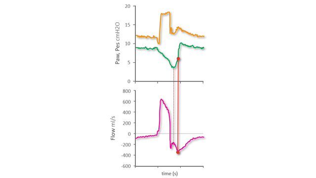
食道压波形上,我们可以看到食道压在最低点后上升的一点,这标志着斜率的明显变化。这一点对应于从同步吸气到随后由延迟切换引起的被动吸气的转变(见下图 4)。

显示长时间指数递减的流量波形
图 3(图像来自 Mojoli et al.Critical Care (2022) 26:32)
显示长时间指数递减的流量波形
图 3(图像来自 Mojoli et al.Critical Care (2022) 26:32)
显示最低点后上升的食道压波形
图 4(图像来自 Mojoli et al.Critical Care (2022) 26:32)
显示最低点后上升的食道压波形
图 4(图像来自 Mojoli et al.Critical Care (2022) 26:32)

较大的不同步 - 自动触发

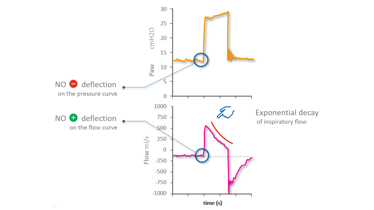
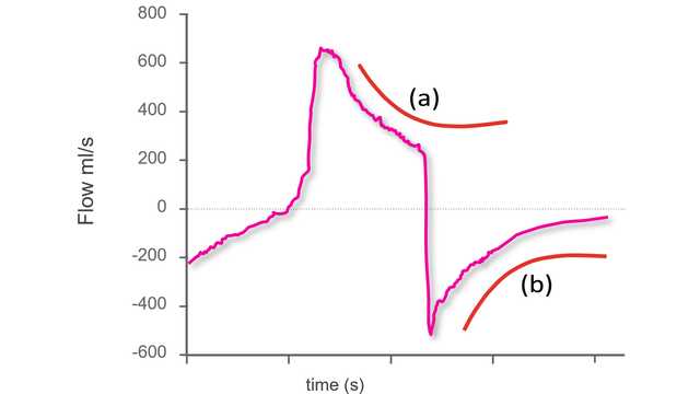
自动触发呼吸是指由呼吸机触发的呼吸,病人无需做出吸气努力。可以通过观察流量和气道压力波形来识别自动触发.没有病人吸气努力的迹象,气道压力没有明显的负偏移,流量也没有明显的正偏移(心脏活动引起的最小振荡除外(见下图 5)。

然而,在吸气流量波形上可以看到自动触发的最强指征,该波形显示一个非常早的峰值,随后是指数递减:整个吸气是由被动吸气导致的。峰值本身的形状也是一个指标:在被动吸气的情况下,峰值是一个尖峰,而在同步吸气时,峰值更弯曲。

与流量和气道压力波形一样,食道压参考波形上没有可见的负偏移,这通常表示吸气努力的开始(见下图 6)。

显示无病人活动的流量和压力波形
图 5(图像来自 Mojoli et al.Critical Care (2022) 26:32)
显示无病人活动的流量和压力波形
图 5(图像来自 Mojoli et al.Critical Care (2022) 26:32)
显示无负偏移的食道压波形
图 6(图像来自 Mojoli et al.Critical Care (2022) 26:32)
显示无负偏移的食道压波形
图 6(图像来自 Mojoli et al.Critical Care (2022) 26:32)

较大的不同步 - 无效吸气努力

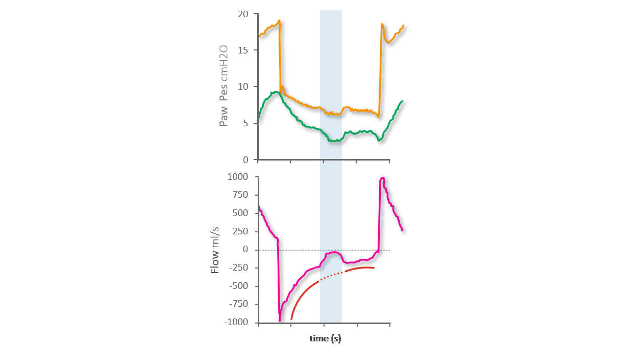
无效的吸气努力是指病人在呼吸机呼气阶段做出的吸气努力,但没有被呼吸机识别,因此也没有得到呼吸机的支持。

流量波形上,您可以将无效的努力识别为暂时影响流量正常指数递减的正偏移。在气道压力波形上,同时发生最小负偏移(见下图 7)。

食道压参考波形上,您可以看到无效吸气努力的开始和结束,显示为食道压的负偏移,影响食道压的正常呼气减少(见下图 8)。

同步活动的流量和压力波形
图 7(图像来自 Mojoli et al.Critical Care (2022) 26:32)
同步活动的流量和压力波形
图 7(图像来自 Mojoli et al.Critical Care (2022) 26:32)
显示影响正常呼气减少的食道压波形
图 8(图像来自 Mojoli et al.Critical Care (2022) 26:32)
显示影响正常呼气减少的食道压波形
图 8(图像来自 Mojoli et al.Critical Care (2022) 26:32)
不同步参考卡

学习发现常见的异步性! 免费参考卡

我们的异步参考卡向您概述了最常见的异步性类型、它们的原因以及如何检测它们。

Timing of inspiratory muscle activity detected from airway pressure and flow during pressure support ventilation: the waveform method.

Mojoli F, Pozzi M, Orlando A, et al. Timing of inspiratory muscle activity detected from airway pressure and flow during pressure support ventilation: the waveform method. Crit Care. 2022;26(1):32. Published 2022 Jan 30. doi:10.1186/s13054-022-03895-4


BACKGROUND

Whether respiratory efforts and their timing can be reliably detected during pressure support ventilation using standard ventilator waveforms is unclear. This would give the opportunity to assess and improve patient-ventilator interaction without the need of special equipment.

METHODS

In 16 patients under invasive pressure support ventilation, flow and pressure waveforms were obtained from proximal sensors and analyzed by three trained physicians and one resident to assess patient's spontaneous activity. A systematic method (the waveform method) based on explicit rules was adopted. Esophageal pressure tracings were analyzed independently and used as reference. Breaths were classified as assisted or auto-triggered, double-triggered or ineffective. For assisted breaths, trigger delay, early and late cycling (minor asynchronies) were diagnosed. The percentage of breaths with major asynchronies (asynchrony index) and total asynchrony time were computed.

RESULTS

Out of 4426 analyzed breaths, 94.1% (70.4-99.4) were assisted, 0.0% (0.0-0.2) auto-triggered and 5.8% (0.4-29.6) ineffective. Asynchrony index was 5.9% (0.6-29.6). Total asynchrony time represented 22.4% (16.3-30.1) of recording time and was mainly due to minor asynchronies. Applying the waveform method resulted in an inter-operator agreement of 0.99 (0.98-0.99); 99.5% of efforts were detected on waveforms and agreement with the reference in detecting major asynchronies was 0.99 (0.98-0.99). Timing of respiratory efforts was accurately detected on waveforms: AUC for trigger delay, cycling delay and early cycling was 0.865 (0.853-0.876), 0.903 (0.892-0.914) and 0.983 (0.970-0.991), respectively.

CONCLUSIONS

Ventilator waveforms can be used alone to reliably assess patient's spontaneous activity and patient-ventilator interaction provided that a systematic method is adopted.

Related articles. Get a deeper look