返回

使用容积二氧化碳图设置 PEEP

文章

作者: Jean-Michel Arnal,高级医师,Ste Musse 医院,Toulon,法国

日期: 23.04.2019

PEEP 用于保持肺通气,防止呼气末肺塌陷。然而,PEEP 可能使正常通气的肺过度扩张,并损害肺灌注。因此,PEEP 的任何变化都可能以不可预测的方式影响整体通气/灌注比值。

使用容积二氧化碳图设置 PEEP

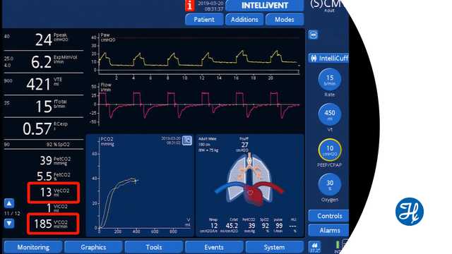
容积二氧化碳图测量每次呼吸呼出的二氧化碳容量 (VeCO2)。PEEP 发生变化后,假设心血管功能和潮气量稳定,VeCO2 的增加意味着整体通气/灌注比值有所改善。相反,VeCO2 的减少意味着通气/灌注比值正在恶化。VeCO2 变化迅速,几分钟后恢复到基线。

这种方法的局限性在于临床医生监测通气/灌注比值的快速变化,例如由于肺过度扩张和肺灌注损伤或改善引起的变化。

PEEP 改变后,肺复张或肺塌陷可能需要更长的时间,并且无法通过该方法进行评估。

观看下面的视频,观看使用 Hamilton Medical 哈美顿医疗公司呼吸机进行通气的演示。 


完整引文如下: (Blankman P, Shono A, Hermans BJ, Wesselius T, Hasan D, Gommers D. Detection of optimal PEEP for equal distribution of tidal volume by volumetric capnography and electrical impedance tomography during decreasing levels of PEEP in post cardiac-surgery patients. Br J Anaesth. 2016;116(6):862-869. doi:10.1093/bja/aew1161​)

Volumetric Capnography: How to set PEEP according to VCO2

Senior intensivist Dr. Jean-Michel Arnal demonstrates how volumetric capnography can help to find the correct PEEP setting.
容积二氧化碳图电子书

免费电子书

不可不知! 容积二氧化碳图的详情

学习如何解读容积二氧化碳图及了解容积二氧化碳图的优点和临床应用。包括自测内容!

Detection of optimal PEEP for equal distribution of tidal volume by volumetric capnography and electrical impedance tomography during decreasing levels of PEEP in post cardiac-surgery patients.

Blankman P, Shono A, Hermans BJ, Wesselius T, Hasan D, Gommers D. Detection of optimal PEEP for equal distribution of tidal volume by volumetric capnography and electrical impedance tomography during decreasing levels of PEEP in post cardiac-surgery patients. Br J Anaesth. 2016;116(6):862-869. doi:10.1093/bja/aew116


BACKGROUND

Homogeneous ventilation is important for prevention of ventilator-induced lung injury. Electrical impedance tomography (EIT) has been used to identify optimal PEEP by detection of homogenous ventilation in non-dependent and dependent lung regions. We aimed to compare the ability of volumetric capnography and EIT in detecting homogenous ventilation between these lung regions.

METHODS

Fifteen mechanically-ventilated patients after cardiac surgery were studied. Ventilator settings were adjusted to volume-controlled mode with a fixed tidal volume (Vt) of 6-8 ml kg(-1) predicted body weight. Different PEEP levels were applied (14 to 0 cm H2O, in steps of 2 cm H2O) and blood gases, Vcap and EIT were measured.

RESULTS

Tidal impedance variation of the non-dependent region was highest at 6 cm H2O PEEP, and decreased significantly at 14 cm H2O PEEP indicating decrease in the fraction of Vt in this region. At 12 cm H2O PEEP, homogenous ventilation was seen between both lung regions. Bohr and Enghoff dead space calculations decreased from a PEEP of 10 cm H2O. Alveolar dead space divided by alveolar Vt decreased at PEEP levels ≤6 cm H2O. The normalized slope of phase III significantly changed at PEEP levels ≤4 cm H2O. Airway dead space was higher at higher PEEP levels and decreased at the lower PEEP levels.

CONCLUSIONS

In postoperative cardiac patients, calculated dead space agreed well with EIT to detect the optimal PEEP for an equal distribution of inspired volume, amongst non-dependent and dependent lung regions. Airway dead space reduces at decreasing PEEP levels.